
Multiple smooth, round, bony overgrowths are seen, and often in both ears.

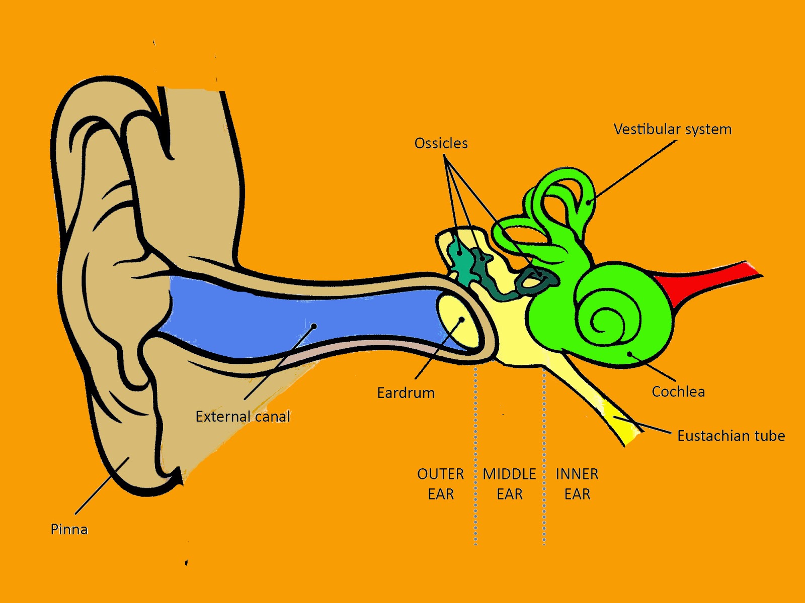
However, these bony overgrowths can also occur in people with no prior water history. The sound waves then travel toward a flexible, oval membrane at the end of the ear canal called the eardrum, or tympanic membrane. It collects sound waves and channels them into the ear canal (external auditory meatus), where the sound is amplified. Defects in the cartilaginous part of the canal, which allow transmission of infection and malignancy, are known as fissures of Santorini. The lateral one-third is bounded by a fibrocartilaginous tube continuous with the auricle 3. An infection caused by a fungus is also diagnosed based on examination or culture (a sample of the pus and debris is grown in a laboratory to identify the. The auricle (pinna) is the visible portion of the outer ear. The external auditory canal is typically 2.5 cm in length and is S-shaped. Physical examination revealed a papillomatous tumor at the posterior wall of the inlet of the left external auditory canal. A 16-year-old female patient presented to our clinic for aural fullness of the left side.
Auditory ear canal skin#
What causes it Exostoses are most commonly found in people with a history of cold water exposure. To a doctor looking into the ear canal through an otoscope (a device for viewing the canal and eardrum), the skin of the canal appears red and swollen and may be littered with pus and debris. This paper reports the first case of fibroepithelial polyp arising independently of the external auditory canal. An otoscope is a hand-held tool with a light and a. A normal eardrum will flex inward and outward in response to the changes in pressure.Children’s Health is proud to become the first pediatric health system in the country to offer Amazon Lockers, self-service kiosks that allow you to pick up your Amazon packages when and where you need them most – 24 hours a day, seven days a week. Exostoses, sometimes called surfers ear, are bony overgrowths in the ear canal. During an ear examination, a tool called an otoscope is used to look at the outer ear canal and eardrum. The external portion of ear canal contains subcutaneous layer, hair follicles, sebaceous glands, and wax-secreting ceruminous glands, between the skin and. It helps the doctor see if there is a problem with the eustachian tube or fluid behind the eardrum ( otitis media with effusion). The auricle is an irregularly concave fibroelastic structure covered by skin that conducts sound waves to the external ear canal. The internal auditory canal (IAC), also referred to as the internal acoustic meatus lies in the temporal bone and exists between the inner ear and posterior cranial fossa. The pinna and the auditory canal are parts of the outer ear. The external ear includes the auricle (or pinna) and the external auditory canal as it leads to the tympanic membrane. The Outer Ear consists of the Pinna, the external auditory meatus (or ear canal) and the tympanic membrane or ear drum. It also shows how well the eardrum moves when the pressure inside the ear canal changes. Its shape functions as a funnel, capturing and channeling sound waves into the auditory canal. Using a pneumatic otoscope lets your doctor see what the eardrum looks like. The doctor will look at each eardrum (tympanic membrane). The doctor will then insert the pointed end (speculum) of the otoscope into the ear and gently move the speculum through the middle of the ear canal to avoid irritating the canal lining. For a baby under 12 months, the ear will be pulled downward and out to straighten the ear canal. Your doctor will gently pull the ear back and slightly up to straighten the ear canal. An ear examination can be done in a doctor's office, a school, or the workplace.įor an ear examination, the doctor uses a special tool called an otoscope to look into the ear canal and see the eardrum.


 0 kommentar(er)
